Lung cancer bronchoscopy biopsy how to do
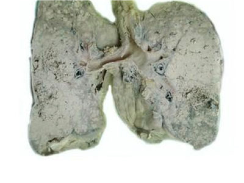
Lung cancer is the most common primary malignant tumor of the lung. The vast majority of lung cancer originates from bronchial mucosal epithelium, so it is also called bronchial lung cancer. In the diagnosis of lung cancer, fiberoptic bronchoscopy is essential. Because lung cancer is a bronchial tumor, especially those early tumors are difficult to find by X-ray or CT film only. At this time, bronchoscopy, which can directly observe the trachea, has become a very effective method. At present, a large number of lung cancer patients are diagnosed by bronchoscopy biopsy or cytology. 
How to do lung cancer bronchoscopy biopsy? Fiberoptic bronchoscopy can be performed under local anesthesia, easy to operate, and less painful for patients. Visible range reaches the main bronchus, leaf bronchus, segment bronchi and secondary bronchi. You can also take tissues for the pathological examination of the lesions you see, or you can use small brushes to brush the cells in the lesions for cytological examination. To achieve a clear diagnosis. After observation through bronchoscopy, the doctor can stage lung cancer according to the extent and severity of the lesion to guide the selection of appropriate treatment options.
Bronchoscopy can not only diagnose lung cancer, but also has a very wide range of uses in treatment. For some intratracheal tumors, bronchoscopy tumor resection can also be performed to avoid the pain of surgery. In addition, for lung cancer patients who are not suitable for surgery, cryotherapy, injection of chemotherapy drugs, photodynamic therapy, internal stent placement, intraluminal radiotherapy, etc. can be performed under bronchoscopy.
Related Articles
- Early symptoms of lung cancer
- 2020-12-17

- Early Signs of Bladder Cancer
- What are the early symptoms of bladder cancer?
- 2020-12-17

- Is metastatic carcinoma easy to metastasize
- Once the cancer has metastasized, it will be very difficult to cure, because many people have lost their lives because of the emergence of cancer, so most people think that cancer is an un
- 2020-08-02

- What does microinfiltrating adenocarcinoma mean?
- Microinfiltrating adenocarcinoma is a type of lung cancer. The reason why it is called microinfiltration means that there is less infiltration around it, which means that it is in the early
- 2020-08-01

- How long can non-small cell adenocarcinoma live
- Adenocarcinoma is one of the most common malignant tumors in the world. Non-small cell adenocarcinoma accounts for about 80% of all adenocarcinomas. About 75% of patients are in the middle
- 2020-08-01

- Hand cancer
- Finger cancer generally refers to the appearance of skin cancer, which is characterized by local cauliflower-like skin and easy bleeding. Finger skin cancer is mostly a malignant tumor that
- 2020-08-01